Information for Peptide: GQVFDVGPR from MK01_HUMAN
1. Biological Peptide
1.1 Native Sequence
R.GQVFDVGPR.Y
1.2 Peptide Properties
| Start | Stop | MW | m/z for 2+ | m/z for 3+ | pI |
|---|---|---|---|---|---|
| 16 | 24 | 973.5 | 487.76 | 325.51 | 5.84 |
1.3 Fragment Ion Table
| AA | b Ion Series | b Ion Mass | y Ion Series | y Ion Mass |
|---|---|---|---|---|
| G | b | y | ||
| Q | b | y | ||
| V | b | y | ||
| F | b | y | ||
| D | b | y | ||
| V | b | y | ||
| G | b | y | ||
| P | b | y | ||
| R | b | y |
2. Synthetic Peptide
2.1 Synthetic (Internal Standard) Peptide Sequence
GQVFDVGP(c5N1)R (Modification: STable isotope added to P8 (13C, 15N) )
2.2 Peptide Properties
| MW | m/z for 2+ | m/z for 3+ | pI |
|---|---|---|---|
| 979.51 | 490.76 | 327.51 | 5.84 |
2.3 Fragment Ion Table
| AA | b Ion Series | b Ion Mass | y Ion Series | y Ion Mass |
|---|---|---|---|---|
| G | b | y | ||
| Q | b | y | ||
| V | b | y | ||
| F | b | y | ||
| D | b | y | ||
| V | b | y | ||
| G | b | y | ||
| P | b | y | ||
| R | b | y |
2.4 Peptide Synthesis Report
 Click to view/download synthesis report for GQVFDVGPR
2.5 QqQ MS/MS
Transition Table:
| Transition | m/z of Transition | Collision Energy |
|---|---|---|
| y8 | 923.48453 | 11.0 |
| y7 | 795.42596 | 17.0 |
| y6 | 696.35754 | 17.0 |
| y5 | 549.28913 | 19.0 |
| y4 | 434.26219 | 25.0 |
| y3 | 335.19377 | 17.0 |
| y2 | 278.17231 | 18.0 |
| b3 | 285.15632 | 15.0 |
| b6 | 646.32009 | 9.0 |
2.6 Calibration Curve of Standard
Description: A dilution series was done on the internal standard peptide from 50 Fmol to 0.1 fmol. Peptide detection was possible to 0.1 fmol.
Experiment Description: Cells from Melanoma cell lineS were lysed in 8M urea/100mM Ammonium bicarbonate buffer. The equivalent of 200,000 cells were loaded onto an SDS gel.
2.7 LC-MRM Analysis of Biological and Standard Peptides Illustrates Elution Times

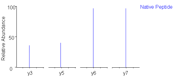
2.8 Pseudo MS/MS Comparison of Transition Patterns for Biological/Standard Peptides